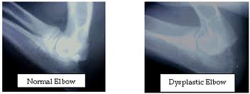
Elbow dysplasia is a general term used to identify an inherited polygenic disease in the elbow of dogs. Three specific etiologies make up this disease and they can occur independently or in conjunction with one another. These etiologies include:
- Pathology involving the medial coronoid of the ulna (FCP)
- Osteochondritis of the medial humeral condyle in the elbow joint (OCD)
- Ununited anconeal process (UAP)
Studies have shown the inherited polygenic traits causing these etiologies are independent of one another. Clinical signs involve lameness which may remain subtle for long periods of time. No one can predict at what age lameness will occur in a dog due to a large number of genetic and environmental factors such as degree of severity of changes, rate of weight gain, amount of exercise, etc. Subtle changes in gait may be characterized by excessive inward deviation of the paw which raises the outside of the paw so that it receives less weight and distributes more mechanical weight on the outside (lateral) aspect of the elbow joint away from the lesions located on the inside of the joint. Range of motion in the elbow is also decreased.
Fractured Coronoid Process (FCP)
Who is usually affected?
-Young dogs of large to giant breeds
-Most frequently affected breeds include Labrador retrievers, Golden retrievers, Rottweilers, and Bernese mountain dogs
What is happening?
-The 3 bones of the elbow joint fit poorly, causing abnormal pressure on the ulna
-A small piece of bone associated with the ulna in the front of the joint (coronoid process) breaks off
-Swelling and pain result from the altered joint mechanics and cartilage destruction
-Arthritis develops
Clinical signs you might notice in your pet
-Limping on a front leg after rest or exercise
-Tiring easily with play
-Resting more than other dogs of similar age and breed, "mellow" puppy
-Head bobbing during walking or running
-Sitting or standing crookedly with a front leg turned outward
Diagnosis
-Careful orthopedic examination to determine which joint(s) are affected
-X-rays are used to evaluate the condition of joint surfaces
-CT scanning may be useful in select cases to evaluate joint surfaces
Surgical treatment
-Removal of the bone fragment and any damaged cartilage (curettage)
-Surgery through small portals (arthroscopy) or a larger incision (arthrotomy) may be indicated
-Surgery on both elbows may be recommended, as the disease is frequently present on both sides
Special postoperative care
-Patient activity is generally limited for 4-6 weeks following surgery, allowing time for joint swelling to subside
Expected results after surgery
-Much initial improvement in the degree of pain and limping
-Ultimate outcome depends on the amount of joint damage present prior to surgery and the degree of elbow misfit ( incongruity )
-Moderate exercise, weight control and medication may be recommended for the long term management of optimal joint health.
Osteochondritis Dissecans (OCD)
Who is usually affected?
-Young dogs of large to giant breeds
What is happening?
-Abnormal maturation of the bone that supports cartilage within joints leads to cartilage thickening, cracking, and exposure of the underlying bone
-Swelling and pain result from the altered joint mechanics and cartilage destruction
-Arthritis develops
-Most commonly affected joints are the shoulder, elbow, knee (stifle), and ankle (hock)
Clinical signs you might notice in your pet
-Limping after rest or exercise
-Tiring easily with play
-Resting more than other dogs of similar age and breed, "mellow" puppy
-Head bobbing during walking or running
-Sitting crookedly with one leg held outward (though this can be normal for puppies)
Diagnosis
-Careful orthopedic examination to determine which joint(s) are affected
-X-rays are used to evaluate the condition of joint surfaces
-X-ray with a contrast liquid (arthrogram) may be needed to fully evaluate the shoulder joint
Surgical treatment
-Removal of the damaged cartilage (curettage)
-Surgery through small portals (arthroscopy) or a larger incision (arthrotomy) may be indicated
Special postoperative care
-Patient activity is generally limited for 4-6 weeks following surgery, allowing time for joint swelling to subside
Expected results after surgery
-Shoulder: Excellent results (pain relief and gradual return to a normal gait) are expected due to the relatively small area of cartilage usually involved and the loose fitting mechanical nature of the joint
-Elbow: Good results are expected unless the area of cartilage involved is very large
-Stifle: Good results are expected if the area of cartilage involved is in a non-weight bearing portion of the stifle; variable results are expected if the area of cartilage involved is in a weight bearing portion of the stifle (additional surgery may be indicated to decrease weight bearing on this site)
-Hock: Fair to poor results (incomplete pain relief and retention of some abnormal gait) are expected due to the relatively large area of cartilage usually involved and the tight fitting mechanical nature of the joint
-Moderate exercise, weight control and medication may be recommended for the long term management of optimal joint health.
Ununited Anconeal Process (UAP)
What is happening?
-The 3 bones of the elbow joint fit poorly, causing abnormal pressure on the ulna
-A small piece of bone associated with the ulna in the back of the joint (anconeal process) fails to attach to or breaks off of the ulna
-Swelling and pain result from the altered joint mechanics and cartilage destruction
-Arthritis develops
Clinical signs you might notice in your pet
-Limping on a front leg after rest or exercise
-Tiring easily with play
-Resting more than other dogs of similar age and breed, "mellow" puppy
-Head bobbing during walking or running
-Sitting or standing crookedly with a front leg turned outward
Diagnosis
-Careful orthopedic examination to determine which
joint(s) are affected
-X-rays are used to evaluate the condition of joint surfaces
Surgical treatment
-Depending on the specifics of each case, recommended surgical options may include:
-Removing the loose piece of bone
-Relieving the pressure on the ulna by cutting the bone (osteotomy) below the elbow joint, encouraging the previously loose piece of bone (anconeal process) to attach to the ulna
Special postoperative care
-Patient activity is generally limited for 4-6 weeks following surgery, allowing time for joint swelling to subside
-Patient activity may be limited for a longer period of time (6-12 weeks) with ulnar osteotomy, allowing time for the anconeal process to fuse with the ulna
Expected results after surgery
-Much initial improvement in the degree of pain and limping
-Ultimate outcome depends on the amount of joint damage present, prior to surgery and the degree of elbow misfit (incongruity)
-Moderate exercise, weight control and medication may be recommended for the long term management of optimal joint health.


